Right Atrium
Update October 29, 2019
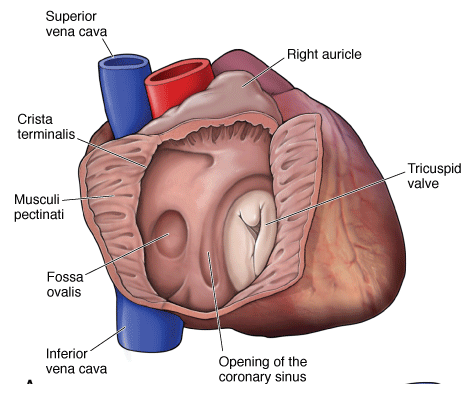
The right atrium receives deoxygenated venous blood from the systemic circulation via the superior and inferior venae cavae and from the coronary circulation via the coronary sinus.
In the fetus, the foramen ovale is an opening in the interatrial septum, which allows blood entering the right atrium from the venae cavae to pass directly to the left side of the heart, bypassing the lungs. Blood flow to the lungs is bypassed because the placenta is responsible for gas exchange in utero. Following birth, the foramen ovale seals shut because of increased pressure in the left side of the heart. Failure of the foramen ovale to seal shut occurs in approximately 20% of the population.
In an adult, this foramen, termed the fossa ovalis, is shut.
On the superior aspect of the right atrium is the auricle, which is an outpouching of tissue derived from the fetal atrium, with rough myocardium on its internal surface known as pectinate muscles. The sinus venarum, which has a smooth wall, is on the internal surface of the remainder of the right atrium. The crista terminalis is the internal vertical ridge that separates the rough portion from the smooth portion of the right atrium. The crista terminalis extends vertically from the superior to the inferior vena cava. The SA node of the conducting system of the heart is located in the superior part of the crista terminalis.